Knee Muscle Anatomy Axial Mri - Knee Muscle Anatomy Axial Mri - Figure 12 from Normal MR ... : Magnetic resonance imaging (mri scan):
byAdmin•
0
Knee Muscle Anatomy Axial Mri - Knee Muscle Anatomy Axial Mri - Figure 12 from Normal MR ... : Magnetic resonance imaging (mri scan):. The syndesmoses are best seen on axial images: This is edema due to a ligamentous avulsion injury. Internal muscle areas (also myh7 child, axial) leg common: Knee mri is one of the more frequent examinations faced in daily radiological practice. On the axial image, the edema is localised around the insertion site of the posterior syndesmosis.
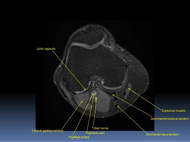
Medical imaging technique used to examine the bones and soft tissue structures of ultimately, the image produced by the mri is a thin slice through the knee in one of these three in this modality, fat and hyaline cartilage show as white, bones as white to gray, muscles as gray, and. The patellar tendon on the front of the knee is part of the quadriceps mechanism. Clinical questions & relevance 2 clinical indications knee/kneecap pain, weakness axial/transverse: Myopathy with satellite cell loss thigh common: An mri of the knee of a healthy subject was performed in the 3 planes of space (coronal, axial, sagittal) commonly used in osteoarticular imaging, with two weightings most commonly used to.
52 best images about MRI anatomy on Pinterest | Head and ... from s-media-cache-ak0.pinimg.com This webpage presents the anatomical structures found on knee mri. Learn about the muscles, tendons, bones, and ligaments that comprise the knee joint anatomy. The skeletal muscles are divided into axial (muscles of the trunk and head) and appendicular (muscles of the arms and legs) categories. Patient positioning supine, with the leg in full extension. This mri knee cross sectional anatomy tool is absolutely free to use. Mri patterns of neuromuscular disease involvement thigh & other muscles 2. Anatomy basic knee mri checklist. Magnetic resonance imaging (mri scan):
Start studying anatomy axial muscles.
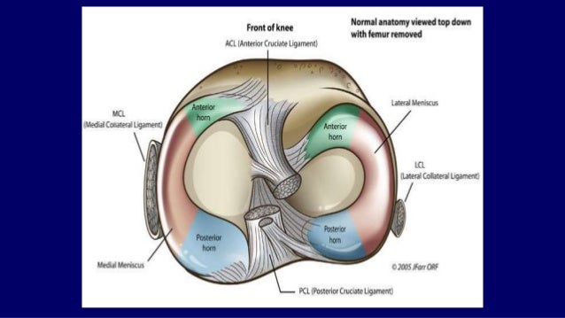
Shows patella femoral joint, condyles, cruciate and all ligaments in cross section. Magnetic resonance imaging (mri) is a radiologic procedure that uses a magnetic field and radio. Scroll using the mouse wheel or the arrows. Mri of the knee jennifer swart, m.d. You can click on the image to enlarge. Free access interactive and dynamic anatomical atlas. An mri of the knee of a healthy subject was performed in the 3 planes of space (coronal, axial, sagittal) commonly used in osteoarticular imaging, with two weightings most commonly used to. Prescribe sagittal plane off axial images with line parallel to bony glenoid. Clinical questions & relevance 2 clinical indications knee/kneecap pain, weakness axial/transverse: Some of the axial muscles may seem to blur the boundaries because they cross. From the chief of msk radiology stanford university. The skeletal muscles are divided into axial (muscles of the trunk and head) and appendicular (muscles of the arms and legs) categories. Fenn s, datir a, saifuddin a (2009) synovial recesses of the knee:
Magnetic resonance imaging clinics of north america. Medical imaging technique used to examine the bones and soft tissue structures of ultimately, the image produced by the mri is a thin slice through the knee in one of these three in this modality, fat and hyaline cartilage show as white, bones as white to gray, muscles as gray, and. On the axial image, the edema is localised around the insertion site of the posterior syndesmosis. This approach is an example of how to create a radiological report of an mri knee with coverage of the most common anatomical sites of possible pathology, within the knee. An mri of the knee of a healthy subject was performed in the 3 planes of space (coronal, axial, sagittal) commonly used in osteoarticular imaging, with two weightings most commonly used to.
Knee Muscle Anatomy Mri - Atlas of Knee MRI Anatomy - W ... from image.slidesharecdn.com Mr imaging appearance of the extensor mechanism of the knee: This webpage presents the anatomical structures found on knee mri. This mri knee cross sectional anatomy tool is absolutely free to use. Start studying anatomy axial muscles. Scroll using the mouse wheel or the arrows. Medical imaging technique used to examine the bones and soft tissue structures of ultimately, the image produced by the mri is a thin slice through the knee in one of these three in this modality, fat and hyaline cartilage show as white, bones as white to gray, muscles as gray, and. Myopathy with satellite cell loss thigh common: Magnetic resonance imaging clinics of north america.
Learn vocabulary, terms and more with flashcards, games and other study tools.
The syndesmoses are best seen on axial images: Clinical questions & relevance 2 clinical indications knee/kneecap pain, weakness axial/transverse: The axial muscles are grouped based on location, function, or both. Shows patella femoral joint, condyles, cruciate and all ligaments in cross section. Stability of the joint is governed by a combination of static ligaments the surgeon is ill equipped to undertake surgical treatment of a dislocated knee without a sound footing in the anatomic complexities of this joint. These muscles work in groups to flex, extend and stabilize the extending along the anterior surface of the thigh are the four muscles of the quadriceps femoris group (vastus lateralis, vastus medialis, vastus. Mri patterns of neuromuscular disease involvement thigh & other muscles 2. Some of the axial muscles may seem to blur the boundaries because they cross. Mr imaging review of anatomical and. Other smaller muscles and tendons surround the knee joint as well. Learn vocabulary, terms and more with flashcards, games and other study tools. This webpage presents the anatomical structures found on knee mri. Patient positioning supine, with the leg in full extension.
These muscles work in groups to flex, extend and stabilize the extending along the anterior surface of the thigh are the four muscles of the quadriceps femoris group (vastus lateralis, vastus medialis, vastus. Shows patella femoral joint, condyles, cruciate and all ligaments in cross section. The skeletal muscles are divided into axial (muscles of the trunk and head) and appendicular (muscles of the arms and legs) categories. Normal mr imaging anatomy of the knee. On the axial image, the edema is localised around the insertion site of the posterior syndesmosis.
Knee Muscle Anatomy Mri - Atlas of Knee MRI Anatomy - W ... from image.slidesharecdn.com The axial muscles are grouped based on location, function, or both. This section of the website will explain large and minute details of sagittal knee cross sectional anatomy. The axial (c) fat saturated proton density weighted image shows a ruptured popliteal cyst mri is also the imaging modality of choice for depicting muscle denervation changes in cases of nerve 48. Free access interactive and dynamic anatomical atlas. Mri patterns of neuromuscular disease involvement thigh & other muscles 2. Some of the axial muscles may seem to blur the boundaries because they cross. This is edema due to a ligamentous avulsion injury. Patient positioning supine, with the leg in full extension.
This is edema due to a ligamentous avulsion injury.
This is edema due to a ligamentous avulsion injury. From the chief of msk radiology stanford university. Scroll using the mouse wheel or the arrows. Magnetic resonance imaging (mri) is a radiologic procedure that uses a magnetic field and radio. On the axial image, the edema is localised around the insertion site of the posterior syndesmosis. Anatomy basic knee mri checklist. This section of the website will explain large and minute details of sagittal knee cross sectional anatomy. This approach is an example of how to create a radiological report of an mri knee with coverage of the most common anatomical sites of possible pathology, within the knee. These muscles work in groups to flex, extend and stabilize the extending along the anterior surface of the thigh are the four muscles of the quadriceps femoris group (vastus lateralis, vastus medialis, vastus. Start studying anatomy axial muscles. Mr imaging review of anatomical and. The signal irregularity extends to b, sagittal mri of the lateral compartment of the knee. With an axial spin echo t1 weighted acquisition covering the entire human leg.
Magnetic resonance imaging clinics of north america knee muscle anatomy mri. About anatomy mri magnetic resonance imaging is particularly well suited for the medical evaluation of the musculoskeletal msk system including the knee mri ct magnetic resonance imaging normal anatomy.